Patient Education
Retinal Detachment
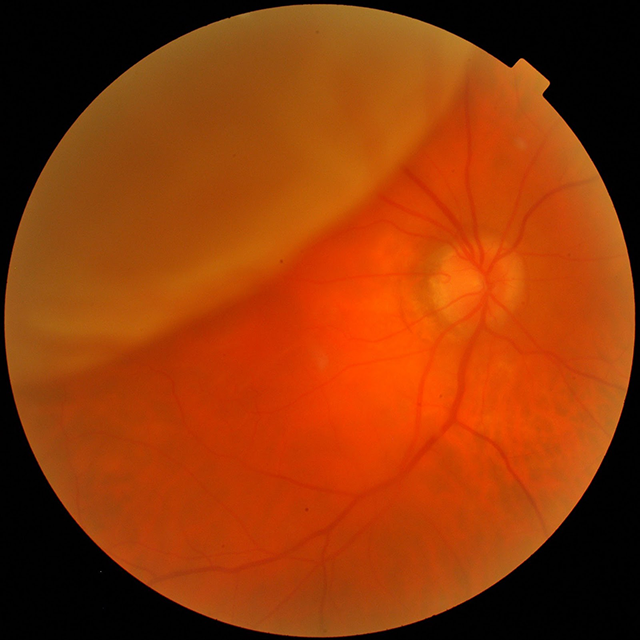
Retinal Detachment
A retinal detachment is a separation of the sensory retina from the retinal pigment epithelium. In most cases, the retina detaches because of small tears or holes that occur as the retina thins with age. This happens because the vitreous (the clear jelly-like substance in the centre of the eye) contracts and becomes more fluid-like with age, and starts to partially separate from the retina (posterior vitreous detachment, PVD). Where the vitreous is firmly attached to the retina, this pulling and separation and may tear the actual retina and cause a detachment.
In about half of the population, the vitreous has separated from the retina (PVD) by age 50. This process can have similar symptoms as a retinal detachment. These symptoms include flashes of light, floaters, shadows across your visual field, and decreased vision.
- Symptoms
- Risk Factors
- Treatment
- Flashes
- Floaters
- Sudden onset of blurred vision
- Shadows or blind spots
- Curtain or veil over your vision
- Extreme nearsightedness (myopia)
- Previous eye surgery
- Previous eye injury
- Previous retinal detachment in one eye
- More common in people over age 50
- A family history of retinal detachment
- Eye disease or inflammation
- Laser surgery
The surgeon directs a laser beam into the eye through the pupil. The laser makes burns around the retinal tear, creating scarring that usually "welds" the retina to underlying tissue.
- Freezing
After giving you a local anesthetic to numb your eye, the surgeon applies a freezing probe to the outer surface of the eye directly over the tear. The freezing causes a scar that helps secure the retina to the eye wall.
- Air or Gas Bubble
The surgeon injects a bubble of air or gas into the center part of the eye (the vitreous cavity). If positioned properly, the bubble pushes the area of the retina containing the hole or holes against the wall of the eye, stopping flow of fluid into the space behind the retina.
- Indenting the surface of your eye
This procedure involves the surgeon sewing (suturing) a piece of silicone material to the white of your eye (sclera) over the affected area. This procedure indents the wall of the eye and relieves some of the force caused by the vitreous tugging on the retina.
- Draining and replacing the fluid in the eye
The surgeon removes the vitreous along with any tissue that is tugging on the retina. Air, gas or silicone oil is then injected into the vitreous space to help flatten the retina. Vitrectomy may be combined with a scleral buckling procedure.